


One of the most serious pathologies of the musculoskeletal system is coxarthrosis of the hip joint. If a visit to a medical facility is delayed, the disease can progress - up to the appearance of acute pain syndrome, which cannot be relieved with analgesics, and the complete loss of motor ability of the joint.
In this article, we will talk in detail about all the nuances related to eliminating the consequences of this pathological process, its stages and preventive procedures.
What is coxarthrosis of the hip joint?
We are talking about a degenerative-dystrophic disease of the hip joint in a severe form, which can provoke a violation of the functional ability of the joint up to its absolute loss. In terms of the frequency of manifestation, coxarthrosis is in second position after deforming arthrosis of the knee joint.
Coxarthrosis of the hip joint is accompanied by degenerative damage to cartilage, the appearance of pathological growths, bone resorption, inflammatory processes and other complications.
That is, this pathology is characterized by damage to the entire joint, which covers cartilage tissue, synovial layer, subchondral bone plate, muscle structures, capsule and ligaments.
The following forms of the disease are also distinguished:
- Primary coxarthrosis. It is considered to be the most common disorder of the hip joint. In older people, this pathology manifests itself against the background of age-related changes;
- Secondary coxarthrosis. Manifests as a result of any disease.
Causes of coxarthrosis
The development of pathology can be provoked by causes of an external, acquired and hereditary nature.
In particular, coxarthrosis can manifest against the background of congenital inferiority of the hip joint, degenerative-dystrophic changes, trauma, inflammatory processes, necrosis of the bone marrow of the femoral head, metabolic disorders, genetic factors, age-related changes, obesity, vascular abnormalities and work in difficult conditions.
It should be noted that almost all joint structures are subject to inflammation.
3 stages of development of coxarthrosis in the hip joint
During the development of the pathological process, the viscosity of the synovial fluid increases, which causes the appearance of microcracks and leads to dehydration of the cartilage surface. This, in turn, contributes to the appearance of crunch and limited mobility. A person feels such unpleasant manifestations during daily stress and physical activity. As the pressure on the lower extremities increases, the exhausted joint adapts to the forced position and begins to destroy nearby structures.
Currently, there are 3 stages of the development of the disease:
- First. Coxarthrosis of the hip joint at this stage has mild symptoms that are inconsistent and appear in the affected area. At the same time, motor activity is preserved, and to relieve pain it is enough to take medicine;
- Other. When a patient is diagnosed with coxarthrosis in the hip joint in stage 1, the disease does not cause much trouble, but when it comes to stage 2 of the disease, the symptoms become more pronounced. The pain becomes more intense and begins to radiate to other parts of the body. The motor ability deteriorates significantly, which becomes especially noticeable after prolonged walking or increased physical exertion;
- Third. If coxarthrosis of the hip joint of the 2nd degree can still be treated, the pathology becomes chronic in the third stage. It is accompanied by constant pain and is transferred to the lower part of the body. The patient loses the ability to move without crutches. In the absence of correct therapeutic measures, atrophy of cartilage and muscle structures occurs.
Types of coxarthrosis
The classification of hip joint pathology is based on one criterion - how the disease arose in the musculoskeletal system. There are two main risk factors that can trigger the onset of the disease – genetic and acquired due to age-related changes. The pathological process is also divided into several types depending on the source of occurrence:
- Primary coxarthrosis. This pathology manifests itself in the hip area and is acquired. In the initial phase, it affects the synovial capsule, after which it passes into the area of the tissues around the joint. Risk factors include increased pressure on the pelvic bones, excessive physical activity, and the presence of inflammatory foci in the lower extremities and spine. Degenerative lesions are concentrated in tissues that have already undergone changes;
- Secondary coxarthrosis. This anomaly is hereditary. It manifests itself in the joints and the musculoskeletal system. The development of the pathological process can begin already in the womb, after a woman has received an injury, as well as against the background of necrosis of the bone marrow of the femoral head.
Types of coxarthrosis due to occurrence:
- Post-infectious. Identified in the presence of consequences after infectious diseases;
- Post-traumatic. Diagnosed in case of complications after limb injury;
- Dyhormonal. Occurs on the background of metabolic disorders or drug overdose;
- Involutive. It appears in people over 50 years old due to the aging of the body.
Diagnostic measures
If you suspect grade 1 or 2 coxarthrosis of the hip joint, you should perform a full body examination before starting treatment. It is also important to have a consultation with an orthopedic doctor who will carry out an examination, give recommendations regarding laboratory tests and prepare an effective treatment plan. Typically, diagnostic measures are limited to the following procedures:
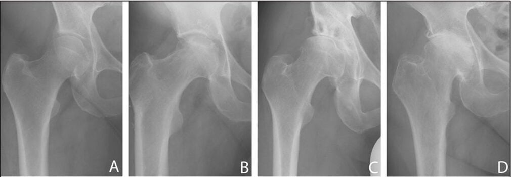
- Radiography. Allows you to study the parameters of the gap between the cartilages, diagnose the presence of pathological growths, and also assess the condition of the femoral head;
- Ultrasound. Makes it possible to trace the etiology of changes in bone structures and ligaments, as well as study the dynamics of the patient's condition and determine the degree of development of the anomaly;
- CT. Allows you to get more detailed information about the condition of the joints and tissues near them;
- MR. This method provides a detailed picture of the condition of all structures in the hip joint.
Treatment of coxarthrosis in the hip joint
If the patient has been diagnosed with coxarthrosis of the hip joint of 1 or 2 degrees, it is possible to achieve effective results through conservative methods. Such therapy is prescribed to the patient individually and covers several techniques, which only together give a positive effect. So, if a patient is diagnosed with coxarthrosis of the hip joint of 1 or 2 degrees and the corresponding symptoms are observed, the following measures can be recommended:
- Use of medication;
- Physiotherapy procedures;
- Shock wave therapy;
- Physiotherapy.
To achieve positive dynamics using conservative methods, the causes that provoked the appearance of coxarthrosis in the hip joint should be eliminated. First of all, you should reduce body weight, which will reduce the load on the joint and minimize the likelihood of further development of the degenerative-dystrophic process.
In addition, you should eliminate the use of tobacco products and increase physical activity, avoid excessive exertion. To prevent the development of pathology, experts recommend the use of orthopedic devices (orthoses and bandages). They allow you to fix the joint and provide the necessary support during physical activity.
Medicine
Medicines are also prescribed on an individual basis. As a rule, patients are advised to take the following medications:

- Non-steroidal anti-inflammatory drugs. These drugs allow you to get a double effect: relieve pain and eliminate the inflammatory process;
- Preparations containing chondroitin, glucosamine and collagen. They allow you to activate restoration processes in cartilage;
- Steroid hormones. Medicines with a strong anti-inflammatory effect. Used in situations where NSAIDs are not significantly effective;
- Muscle relaxants. Medicines that relieve muscle tone, which is a necessary condition for relieving pain of increased intensity;
- Means that normalizes blood circulationand improving the trophism of tissues located near the joint;
- Vitamin B. Complexes containing this vitamin are prescribed to improve nerve transmission, which is of particular importance when the ends are compressed by affected structures.
In case of pain of considerable intensity, it is also recommended to perform periarticular blockades. They are only performed under the supervision of professional specialists in a clinical setting. In this case, special solutions with steroid hormones and anesthetics are injected into the joint.
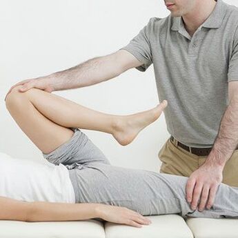
Gymnastics for coxarthrosis in the hip joint
Particularly effective in restoring motor function and reducing muscle spasms are special exercises recommended for coxarthrosis in the hip joint. Due to the optimally selected load, it is possible to relieve pain and increase the range of motion. In addition, a correctly composed complex allows you to prevent atrophic processes in the muscles and relieve spasms if pinched nerve endings are observed against the background of the disease.

Gymnastics for coxarthrosis of the hip joint also helps improve blood flow in the affected area and allows you to speed up recovery processes.
When choosing exercises, the specialist must take into account the destruction of the hip joint and the physical condition of the patient.
Physiotherapy methods
Massage procedures and physiotherapy can have a particularly pain-relieving, anti-inflammatory and relaxing effect. They also help maintain muscle tone in the limbs, which prevents atrophic processes.
For abnormalities of the hip joint, the following procedures are performed:
- UHF;
- Laser exposure;
- Ultrasound treatment;
- Magnetotherapy;
- Exposure to direct current in combination with medication;
- Paraffin treatment;
- Phonophoresis.
The above treatment will only give a positive effect if the patient has been diagnosed with coxarthrosis in the primary stages.
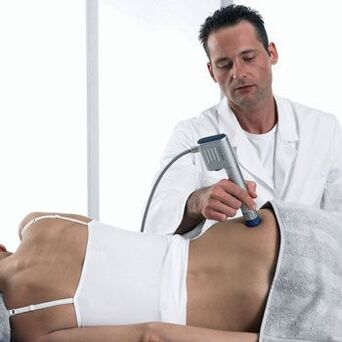
Shock wave treatment for coxarthrosis
For coxarthrosis of the first or second stage, shock wave therapy provides significant positive dynamics. For example, a course of 10-15 shock wave treatment procedures can reduce the negative manifestations characteristic of stage 2 pathology to signs of the initial stage of the disease.
It is important to understand that only timely treatment sessions can provide the best recovery effect. At the same time, it will be possible to reduce the number of SWT procedures.
But the most important positive aspect when affecting the affected joint with shock waves is the ability to normalize blood circulation, which facilitates the accelerated supply of important nutrients involved in regenerative processes to various structures of the hip joint.
In addition, as part of the implementation of shock wave therapy, it is possible to crush pathological bone growths, which contribute to significant irritation of articular tissue and prevent regeneration.
Physiotherapists and neurologists with professional experience operate in the clinics. They are fluent in working with the latest physiotherapeutic methods, which include the shockwave method. In addition, specialists have the ability to work with modern equipment. This gives a guaranteed positive effect and allows you to shorten the treatment period.
Surgery
Unfortunately, many patients delay contacting a medical facility and only see a specialist when irreversible processes begin to occur in the hip joint.
For advanced third or fourth stages of the disease, the only effective method is surgery. It will restore motor ability and eliminate acute pain, that is, significantly improve the patient's quality of life.
As a rule, surgery is prescribed in the following situations:
- Painful sensations of increased intensity that cannot be relieved by medication;
- Lack of interarticular space;
- Violation of the integrity of the femoral neck;
- Significant limitation of physical activity.
Given the intensity of joint damage and changes in bone tissue, patients may be prescribed the following types of interventions:
- Arthrodesis. An intervention that creates complete immobility of the joint. For this purpose, special metal plates are used;
- Osteotomy. A surgical procedure consisting of an artificial fracture of the femur to straighten its axis. The resulting parts are placed in the most optimal position, which allows you to remove excessive load from the affected joint;
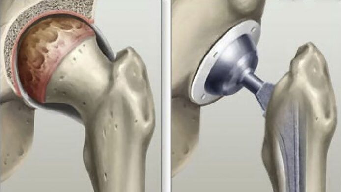
- Arthroplasty. The only method through which it is possible to restore all the functionality of the hip joint and achieve a complete recovery of the patient. After using this method to eliminate coxarthrosis, a person forgets about problems with joints for 20-30 years.
Medical centers perform surgical procedures in the area of the hip joint of any complexity. They are performed by highly qualified specialists using modern tools and technology, which eliminate any errors during the intervention.
Complications of the disease
When the pathological process is at an advanced stage, joint mobility is significantly limited, a person loses the ability to walk and care for himself, and pathological tissue fusion is observed. Moreover, such an anomaly can have an undesirable effect on the way, which is caused by the appearance of lameness and a decrease in the size of the limb.
Preventive actions
Patients with pain in the hip joint should be observed by a specialist and use special orthopedic devices when performing work and physical activity. In addition, after the operation, it is necessary to undergo radiography 3 times a year to monitor the condition of the joint.