


Thoracic osteochondrosis is a chronic disease of the spine in which degenerative-dystrophic changes occur in the intervertebral discs.
The thoracic spine is less often affected by osteochondrosis compared to the cervical and lumbar spine. This is explained by the fact that it is relatively inactive, stable and well strengthened by a muscular corset. Even more rare are its complications - protrusion and herniated disc.
However, this disease presents extensive symptoms that significantly reduce the quality of life and therefore requires treatment. The use of medication only alleviates the symptoms and provides a temporary effect that does not affect the development of the disease.
To reliably eliminate symptoms, you need to affect the cause of the development of degenerative processes in the discs. For this purpose, the clinic uses complex therapy, which gives positive results in more than 90% of cases. It includes methods of oriental reflexology and physiotherapy - acupressure, acupuncture, moxotherapy and other therapeutic procedures.
Symptoms, signs
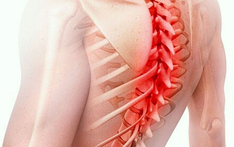
In osteochondrosis, a flattening of the intervertebral discs occurs and the vertebrae come together, which leads to compression of the spinal nerve roots. This causes pain between the shoulder blades (usually described as a stab).
Pain syndrome in thoracic osteochondrosis can be acute, intense or chronic, moderate.
In the first case, acute pain occurs suddenly and is called dorsago. In the second case, the pain is felt constantly, has an aching character and is called dorsalgia.
Irritation from a pinched root spreads along the nerve, radiates into the chest and becomes the cause of intercostal neuralgia - stabbing, cutting or burning pain in the chest, which is intensified by breathing, moving, coughing, sneezing, laughing.
Another characteristic symptom of thoracic osteochondrosis is pain in the heart area, which is accompanied by signs of cardioneurosis - palpitations, palpitations, increased heart rate.
Pinched nerve root leads to disruption of innervation, numbness, weakness in the hand, a feeling of coldness in the hand, cyanosis (blue discoloration) or blanching of the skin. These symptoms are usually unilateral.
Pain with osteochondrosis can also radiate to the shoulder, under the shoulder blade and to the forearm.
Other symptoms of the disease are stiffness, tension in the back, numbness in the paravertebral region, shoulders, neck collar area, difficulty breathing, feeling of a lump in the chest.
The nerves that arise from the spinal cord in the thoracic region play an important role in innervating the entire body. Therefore, symptoms of osteochondrosis can appear in areas that are apparently not related to the spine. For this reason it is called the "chameleon disease".
These symptoms include:
- heartburn, bloating,
- loss of appetite, nausea,
- indigestion (dyspepsia),
- cough,
- cold feet,
- numbness in the body,
- pain in the right hypochondrium,
- stomach discomfort,
- sweating
In addition, thoracic osteochondrosis is manifested by reduced blood supply to the brain - headaches, instability of pressure, dizziness, unsteady gait and loss of coordination.
Causes of development, stages
The main role in the development of the disease is played by muscle spasms and tension (hypertonicity) in the back muscles. These spasms occur during a sedentary lifestyle, poor posture or prolonged stay in a static, uncomfortable position (for example, at an office desk or while driving).
On the other hand, monotonous, hard physical work also provokes the appearance of persistent muscle spasms in the back (for example, work with raised arms).
Muscle spasms inhibit circulation and inhibit blood flow to the spine. Because of this, the nutrition of the intervertebral discs deteriorates.
Intervertebral discs are shock-absorbing cushions of connective tissue found between the vertebrae. In the center of each slice is a pulpy, semi-liquid core that contains a lot of moisture. Water provides resistance to loads and resistance to compression.
Along the outer circumference of each disc is reinforced with a stiff fibrous ring. The connective tissue of the discs consists mainly of collagen - this substance is synthesized in the body and must be constantly supplied to the joints, intervertebral discs and other connective tissue, cartilage tissue for their continuous regeneration.
Muscle spasms disrupt blood flow, resulting in not enough collagen reaching the discs for normal tissue repair. Lack of oxygen leads to a slowdown in metabolic processes.
As a result of metabolic disturbances, the tissue renewal of the intervertebral discs is slowed down and their wear accelerates. This leads to dystrophy and degenerative changes - the discs become dehydrated, crack, dry out, flatten and lose their shock-absorbing properties and elasticity.
Back muscle spasms are the main cause of excessive stress on the spine in the thoracic region. If the intervertebral discs in the cervical region are pressed by the weight of the head, which increases with incorrect posture, and the lower back is pressed by the body weight, which increases with excess weight, then muscle spasms in the thoracic region play an exceptional role in the development of the disease. These spasms not only inhibit blood flow, but also tighten the spine and compress the intervertebral discs both during the day and at night. Intervertebral discs are practically deprived of the possibility not only of cellular renewal, but also of simple rest and recovery. Therefore, the first thing a doctor should do when treating thoracic osteochondrosis is to relax tense back muscles, eliminate muscle spasms and hypertonicity. Without this, effective treatment of the disease is impossible.
The flattening of the intervertebral discs leads to the spaces between the vertebrae becoming smaller, the vertebrae coming closer to each other and pinching the nerve roots. This causes pain, which causes a reflex muscle spasm and further increases the pressure on the discs. Therefore, with the appearance of pain, the development of the disease, as a rule, accelerates.
These degenerative-dystrophic changes correspond to the first stage of osteochondrosis.
Important!
In old age, thoracic osteochondrosis usually develops against the background of general dehydration and metabolic disturbances in the body. This is especially manifested by a decrease in height in older people, which occurs due to thinning of the intervertebral discs.
In the second stage, the outer fibrous ring becomes unfibrous. Its tissue becomes loose, weakened, and it cannot sustain the internal load. As a result, a protrusion of the disk (usually local) occurs in the form of a protrusion.
A projection directed towards the spinal cord is called dorsal. Projections directed to the side are called laterals. The rarest case is uniform protrusion of the disc along the entire circumference.
The appearance of protrusions usually leads to increased pain. An X-ray clearly shows a decrease in the height of the distance between the vertebrae as well as the development of osteophytes - bone growths. They are formed along the edges of the vertebrae to compensate for the loads on the spine as the intervertebral discs cope with them less and less.
In the third stage of the disease, the fibrous ring of the disc cannot withstand internal pressure and rupture. Through the resulting space, part of the nucleus pulposus of the disc is pushed out - an intervertebral hernia occurs.
At the fourth stage of the disease, the range of motion in the back decreases sharply, the pain syndrome becomes constant, and a comprehensive picture of neurological disorders develops.
Diagnostics
At the first appointment, the doctor asks the patient about the symptoms, the circumstances of their occurrence, studies the medical history, conducts an external examination, pays attention to posture, the presence or absence of spinal deformities (scoliosis, kyphosis).
The cause of pain syndrome (dorsago, dorsalgia) can be both osteochondrosis and vertebral displacement (spondylolisthesis), ankylosing spondyloarthrosis, ankylosing spondyloarthrosis.
Osteochondrosis of the thoracic region is usually accompanied by muscle tension in the back and hypertonia of the spinal muscles. The doctor performs palpation and uses successive pressures to find pain (trigger) points that correspond to the centers of muscle spasms.
To get more detailed information, the doctor prescribes an X-ray or MRI.
X-rays for thoracic osteochondrosis provide the most general information - it helps to distinguish the disease from spondylolisthesis, to see osteophytes and narrowing of the gaps between the vertebrae.
Magnetic resonance imaging shows better soft connective tissue. With its help, the doctor can examine in detail the structure of the intervertebral discs, see the protrusion, hernia (its size, location, shape), as well as the condition of ligaments, intervertebral joints, blood vessels, nerve roots and see spinal stenosis (or its danger).
Based on the MRI data, the doctor makes a diagnosis and determines an individual treatment plan.
Treatment of osteochondrosis in the thoracic region
Drug treatments
To relieve pain in the back and intercostal neuralgia in thoracic osteochondrosis, non-steroidal anti-inflammatory drugs in the form of ointments, tablets or injections can be used. The main effect of these drugs is anti-inflammatory, so their use is justified in cases where a pinched nerve root is accompanied by its inflammation, that is, with thoracic radiculitis. NSAIDs also reduce inflammation in muscle tissue on the background of spasms and persistent hypertension.
In case of acute pain syndrome, paravertebral or epidural blockade can be used - an injection of an analgesic. In the first case, the injection is made at the place where the nerve root is pinched, in the second case, in the area between the periosteum of the vertebra and the membrane of the spinal cord.
To relieve muscle tension and reduce the pressure on nerve roots, blood vessels and intervertebral discs, muscle relaxants and antispasmodics are used.
Vitamin complexes are prescribed to nourish nerve tissue and prevent their atrophy.
To slow down the process of destruction of connective tissue, chondroprotectors can be prescribed.
These drugs have a symptomatic effect and can slow down the development of the disease somewhat, but in general they have almost no effect on the process of degenerative changes in the intervertebral discs.
Non-medical treatment
Non-drug treatment of thoracic osteochondrosis includes methods of physiotherapy, reflexology and physiotherapy.
The main goals of the treatment are relief of the inflammatory process, improvement of blood circulation and restoration of metabolic processes in the spinal discs, stimulation of cellular renewal of connective tissue. The clinic uses complex therapy using oriental medicine methods for this purpose.
Important!
Physiotherapy exercises help to form and strengthen the muscle corset, eliminate irrational loads on the spine and serve as a prevention of overload and the formation of muscle spasms.
Surgery
For large hernias, especially dorsal ones, with the threat of spinal stenosis, and especially if present, a surgical operation - discectomy - may be indicated.
Part of the disc is removed, or the entire disc is removed and replaced with a prosthesis. Despite the fact that discectomy is a common type of surgical procedure, operations on the thoracic region are performed extremely rarely.
Treatment in the clinic
Treatment of thoracic osteochondrosis in the clinic is carried out in complex sessions, which include several procedures - acupuncture, acupressure, moxotherapy, stone therapy, vacuum therapy, hirudotherapy for individual indications.
High efficiency is achieved due to the synergy between individual methods and elimination of the cause of the disease.
- Acupressure. By pressing strongly on the trigger points on the back, the doctor eliminates muscle spasms, tension, overload, improves blood circulation and restores unobstructed blood flow to the spine. Thanks to this, the load on the intervertebral discs is reduced, and the processes of metabolism and tissue regeneration are accelerated when the influx of oxygen and collagen increases.
- Acupuncture. Insertion of needles into bioactive points on the back, legs, arms, head, chest eliminates symptoms associated with decreased innervation - numbness, weakness in the arm. With the help of this procedure, intercostal neuralgia and other vertebrogenic pains are relieved. In addition, acupuncture enhances the effect of acupressure and is anti-inflammatory and anti-edematous.
- Moxibustion therapy. Heating bioactive points in the spine is done with a smoldering wormwood cigar. This procedure activates metabolic processes, increases blood flow to the intervertebral discs, stimulates and accelerates their recovery.
- Vacuum therapy. Cupping massage and cupping create blood flow and help improve blood circulation.
- Manual therapy. Using a gentle traction on the spine, the doctor relieves the intervertebral discs, increases the distance between the vertebrae, releases compressed nerve roots, relieves pain and increases the range of motion in the back.
Gentle traction, or traction, is the only manual therapy technique indicated for thoracic osteochondrosis. Before you start, the doctor must thoroughly relax the back muscles, eliminate spasms and free the spine. To do this, the muscles are well warmed up and relaxed through massage. If this is not done, the application of physical exertion can lead to injury - fracture, sprain or fracture. Hardware methods of spinal traction for osteochondrosis are ineffective and even dangerous, so they are not used in the clinic.
Hirudotherapy
The placement of medicinal leeches improves local blood circulation, blood supply to the intervertebral discs and has an anti-inflammatory effect.
Stone therapy
Smooth stones heated to a certain temperature are placed along the spine to warm and relax the back muscles, improve blood circulation and stimulate blood flow.
The duration of a treatment session in the clinic is 1-1. 5 hours depending on individual indications. The course of treatment usually includes 10-15 complex sessions. After completion, a follow-up MRI is performed to evaluate the treatment results achieved.
Complications
The main complication of thoracic osteochondrosis is spinal stenosis due to disc herniation with the development of body paralysis.
Other possible complications are associated with disruption of the innervation of the body due to pinching of the spinal nerve roots: development of diseases of the gastrointestinal tract, kidneys, heart and reproductive system.
Prevention
To prevent the development of thoracic osteochondrosis, you should avoid a sedentary lifestyle and monitor your posture.
Important!
If a child or teenager has scoliosis, it is advisable to cure this disease without hoping that it will go away on its own. Lateral curvature of the spine occurs as a growing pain but can last a lifetime.
In this case, persistent muscle tension and spasms will be inevitable, which in turn will lead to the development of osteochondrosis and possibly its complications. And this is in addition to the fact that scoliosis itself is fraught with complications from the respiratory, digestive and cardiovascular systems.